Description:
| Courtesy Meyer
Laboratory |
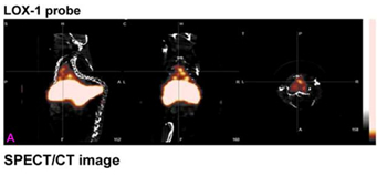
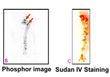
| SPECT/CT imaging (A), phosphor
imaging (B) and Sudan IV staining (C). All mice injected with LOX-1 probe
had hotspots in the aortic arch (sagittal, coronal and transversal
plane). |
Executive Summary
University of Virginia researchers have
developed a novel, non-invasive imaging technique for the detection and
assessment of atherosclerotic plaques. This technique detects atherosclerotic
lesions and can identify which are rupture-prone, vulnerable plaques, leading to
an accurate assessment of heart disease and helping to guide physicians to an
effective treatment plan. Despite a tremendous clinical need, no other method
currently exists that can provide this type of
information.
Advantages
This novel technique offers
the following advantages:
- Provides non-invasive, multi-modality imaging of atherosclerotic plaques
for accurate diagnosis of the state of cardiovascular disease
- Can differentiate between stable and vulnerable plaques, allowing for
targeted medical intervention before a rupture event that could cause heart
attack or stroke
- Imaging is consistent and reliable because it is targeted towards a well
characterized molecular marker of atherosclerosis, LOX-1
Background
Atherosclerosis, characterized by the
thickening of artery walls, is a major cause of many cardiovascular disease
states, including myocardial ischemia, acute myocardial infarction and stroke.
Unfortunately, there are currently no established non-invasive methods for
identifying the rupture-prone atherosclerotic plaque in the living animal. The
LOX-1 receptor has been shown to play a critical role in atherogenesis and the
vulnerability of established plaques.
The U.Va. inventors of this
technology are leading experts in the clinical aspects of atherosclerosis,
cardiovascular magnetic resonance imaging, and the targeted delivery of imaging
and therapeutic agents.
Technical Description
Craig
H. Meyer, Ph.D., and colleagues have developed a novel, non-invasive imaging
probe targeted to LOX-1. In a murine model of atherosclerosis (Apo
E%u207b/%u207b), the probe has been demonstrated to bind to lesions
in vivo and used to detect and assess atherosclerotic plaque using
hybrid SPECT/CT and MRI. After 24 hours, the probe was fully cleared in
wild-type mice and 70–80 percent cleared in Apo E%u207b/%u207b mice.
Results were confirmed by ex vivo fluorescence
imaging.